The Revolution of Surgical Guides: 3 Case Studies Worth Reading
The application of 3D-printed surgical guides in veterinary medicine has brought about a pioneering change: here we present 3 very illustrative examples.

Problems of the patella primarily occur in small dogs but we have also created a trochlear cap for a 50 kg Akita at LimesVet. In many cases, the groove on the lower end of the femur is so worn (or underdeveloped) that the patella dislocates medially or laterally. Besides, another type of congenital abnormality is that the patella gets above the groove (where the cartilaginous edge no longer extends) when the limb is extended, so the patella dislocates because it does not move in its normal groove. The most common sign of patellar luxation is that the dog picks up its leg for shorter or longer periods without putting any weight on it while running, and this movement disorder appears more and more over time. If it is not treated properly, it can even become permanently fixed in this abnormal position (which results in the shortening of muscles and ligaments in addition to a continuous limp).
These problems are generally doctored with conventional operations during which the groove on the femur is deepened, so the patella can move in this new trench. In some cases, however, arthrosis develops over time after such a surgery. In other words, the cartilage that covers the bone wears away (and bone spurs - so-called exostoses - appear). This may require further operations and regular physiotherapy is also recommended.
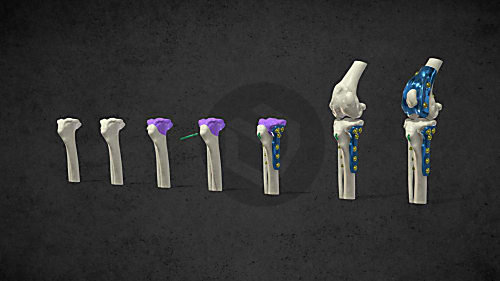
Another widespread solution is that a serially manufactured implant is surgically placed in the dog’s knee. In this case, you can choose from standard sizes, that is, the implant does not fully reflect the internal build-up of the animal. During such an operation a piece is cut from the trochlea of the femur (which hosts the patellar groove) together with the bone and the cartilage (this process is termed osteotomy). Then, the implant is attached onto the smooth surface formed. Therefore, it can be considered an invasive method.
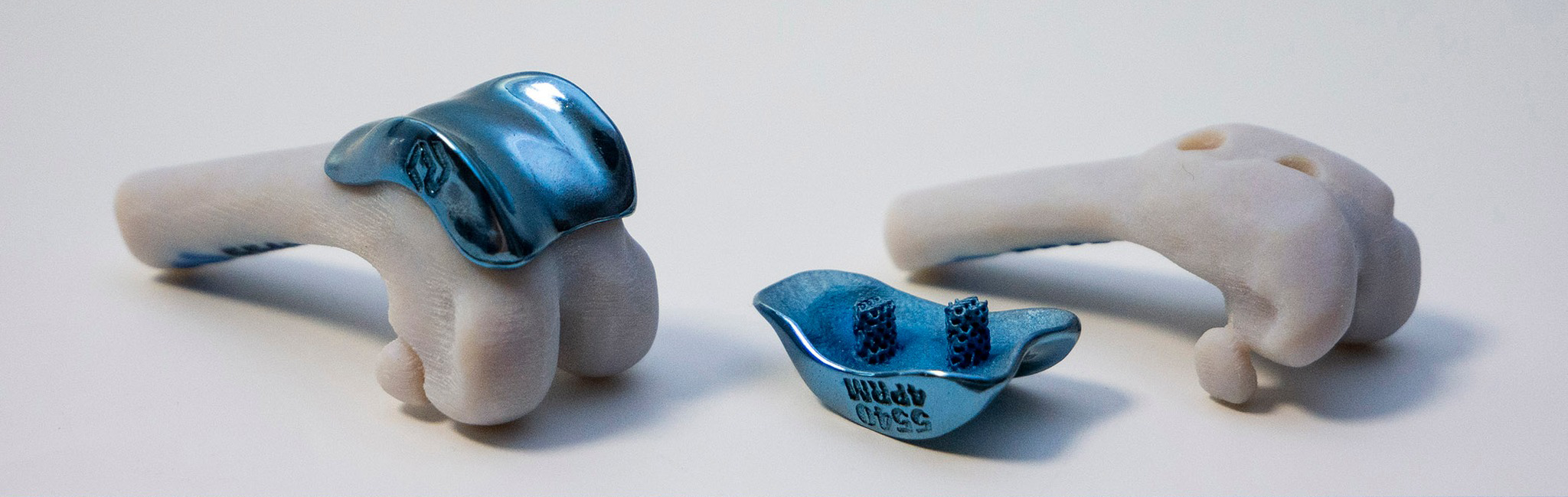
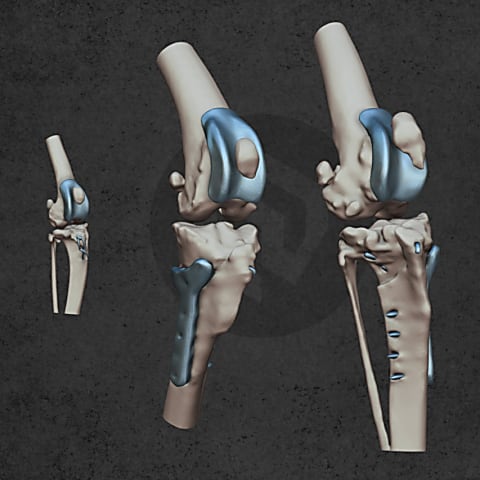
Our uniquely designed TROCA 55 Trochlear Cap covers the required bone and cartilage surfaces like a vault. We always custom design our prostheses and 3D print them using a special alloy of titanium. This minimally invasive procedure requires veterinarians to make holes only at the attachment points - these bores are created with the help of drilling guides tailored to the unique anatomy of the individual dog. As a result of natural ossification, the bone will grow into the latticed pin over time and provide additional stabilization to the implant. Veterinarians can use one of the drilling guides we provide for practice and demonstration to the owner, while the other can be used during the operation. The set also contains two printed anatomical models. One of them shows the femur before and the other after drilling (the metal implant can also be tried on the latter).
Since this implant fits the anatomy of the given animal as much as possible, it is beneficial in terms of biomechanics and it significantly reduces the rehabilitation period.

The application of 3D-printed surgical guides in veterinary medicine has brought about a pioneering change: here we present 3 very illustrative examples.
Today it is obvious that the appearance of 3D technology in the field of medicine opens new horizons for medical professionals in treating diseases. Currently we are just getting to know the advantages of this technology. However, thanks to the rapid development in this field, the question arises as to how to integrate all these achievements into the daily healing practice.
The combined application of custom 3D-printed and traditional implants represents a significant advancement in treating complex orthopedic conditions.

Patient-specific surgical equipment

Personalized implants
Personalized implants

Patient-specific surgical equipment