When a dog is diagnosed with patellar luxation, the conversation quickly turns to the groove - specifically, whether it's deep enough to keep the patella in place. That instinct isn't wrong, but it's incomplete. And in surgery, incomplete thinking leads to incomplete results.
The most common misconception in managing patellar luxation is this: fix the groove, fix the luxation. It's an appealing idea - simple, anatomically intuitive, and easy to explain in a consulting room. But it oversimplifies a condition that is, in most cases, a syndrome rather than a single lesion.
When the groove alone is addressed without evaluating the rest of the limb, the consequences can include:
- Recurrence of luxation, sometimes within months of surgery;
- Persistent lameness despite a technically sound trochleoplasty;
- Progressive osteoarthritis, accelerated by continued mechanical malalignment;
- Inconsistent outcomes that are difficult to predict or explain.
The goal of this summary is to reframe the way we think about patellar luxation - for those who are trying to understand what their pet is facing, and for clinicians planning treatment. The groove matters. But it is one component of a much larger mechanical story.
Section 1: What Is patellar luxation, really?
Patellar luxation means the patella (the kneecap) slips out of its normal position in the trochlear groove of the femur. Mechanically, the patella acts as a pulley - it amplifies the force of the quadriceps muscle and transfers it through the patellar tendon to the tibial tuberosity, extending the stifle (knee) joint.
For that system to work, the patella needs to track cleanly through the groove through the full range of motion. When it doesn't - when it drifts medially (inward) or laterally (outward) - you get luxation.
In practice, this shows up as:
- Intermittent or persistent lameness, often described as "skipping";
- Bunny-hopping gait in bilateral cases;
- Stiffness, reduced range of motion, or reluctance to jump;
- In chronic cases, joint pain and progressive arthritis.
Luxations are graded from I (occasional, manually reducible) to IV (permanent, irreducible), and both medial and lateral forms exist - though medial luxation is far more common in small and toy breeds, while lateral luxation is more typical in large breeds.
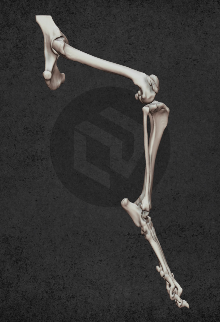
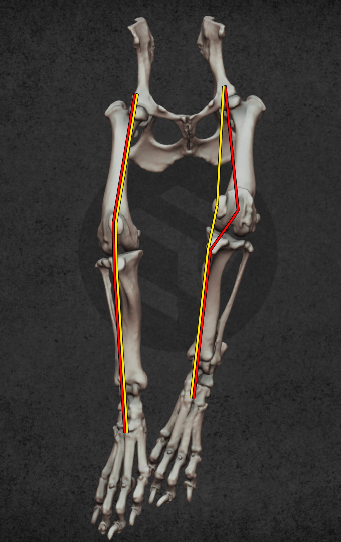
The critical point is this: patellar luxation is rarely caused by a bad groove alone. It is driven by the geometry and alignment of the entire limb.
Section 2: The big picture - Patellar luxation as a limb alignment disorder
Think of the extensor mechanism - the quadriceps muscle, patella, patellar tendon, and tibial tuberosity - as a cable running through a system of pulleys. Every component of that system, from the femur down to the foot, influences the direction of pull.
Patellar luxation is what happens when the cable is being pulled off-centre by some combination of:
- Femoral changes: rotational or angular deformities that aim the quadriceps mechanism medially or laterally;
- Tibial changes: torsion or deviation of the tibia that repositions the tibial tuberosity out of the ideal line of pull;
- Soft tissue changes: chronic luxation creates contractures on one side and laxity on the other, actively perpetuating maltracking;
- Trochlear changes: a shallow or poorly shaped groove that fails to guide the patella.
None of these factors exists in isolation. They interact. And that is why treatment planning must account for all of them.
Section 3: Preoperative assessment - The whole-limb checklist
Good surgical outcomes begin before the first incision. A thorough preoperative evaluation should answer one central question: where is the extensor mechanism being pulled off-track, and why?
Clinical examination
- Luxation grade, reducibility, presence of crepitus;
- Pain response and range of motion assessment;
- Limb alignment observed both in stance and during gait;
- Direction of patellar displacement through flexion and extension.
Imaging
Plain radiographs are a starting point, but a properly planned CT scan with objective measurements is the gold standard for surgical decision-making. CT allows accurate assessment of femoral and tibial torsion, angular deformities, and the spatial relationship of the tibial tuberosity to the trochlear groove - information that is simply not visible on X-ray.
With a clear picture of the limb mechanics, the surgical plan becomes a targeted correction rather than a generic procedure.
Section 4: The four pillars of surgical correction
Pillar 1. Osseous Reconstruction: correcting what drives maltracking
The bones set the trajectory of the entire extensor mechanism. If the femur is internally rotated or the distal femur has angular deformity, the quadriceps is pulling the patella off course before it even reaches the groove. No amount of groove deepening will compensate for that.
Femoral deformities to consider include:
- Rotational deformity (excessive internal or external torsion);
- Varus or valgus angulation;
- Distal femoral shape contributing to persistent medial or lateral quadriceps pull.
If femoral malalignment is identified as a primary driver, corrective osteotomy should be part of the plan. This is not optional if the deformity is significant - groove work alone cannot redirect a misdirected mechanism.
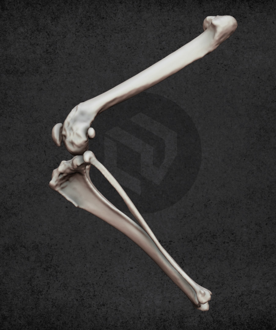
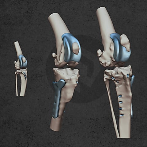
Pillar 2. Tibial Tuberosity Transposition: realigning the line of pull
The tibial tuberosity is where the patellar tendon inserts - it is the distal anchor of the entire extensor mechanism. Its position relative to the trochlear groove determines the angle at which the patella is being pulled.
Tibial tuberosity transposition (TTT) repositions this anchor so that the patellar tendon pulls the patella centrally through the groove. It is not a supplementary step - in most cases of significant luxation, TTT is essential for durable correction.
A corrected groove can still fail if the tibial tuberosity is pulling the patella back off course. The mechanical axis of the limb must be addressed as a whole.
Pillar 3. Soft Tissue Balancing: restoring tension equilibrium
Chronic patellar luxation is not just a bone problem. Over time, soft tissues adapt - the structures on the side toward which the patella is displaced become contracted and shortened, while those on the opposite side become overstretched and lax.
These adaptations actively maintain the maltracking even after bony correction. Addressing them is not optional.
The principles of soft tissue balancing are:
- Release structures that are abnormally tight and pulling the patella off track - this may include the retinaculum, joint capsule, and fascial layers;
- Imbricate (reef) structures that are overstretched and failing to provide medial or lateral support;
- Reassess tension through the full range of motion, not just at a single joint angle;
- Consider quadriceps contracture in chronic or severe cases, where the quadriceps muscle itself may require attention.
Soft tissue balancing is what transforms a mechanically corrected limb into a clinically functional one.
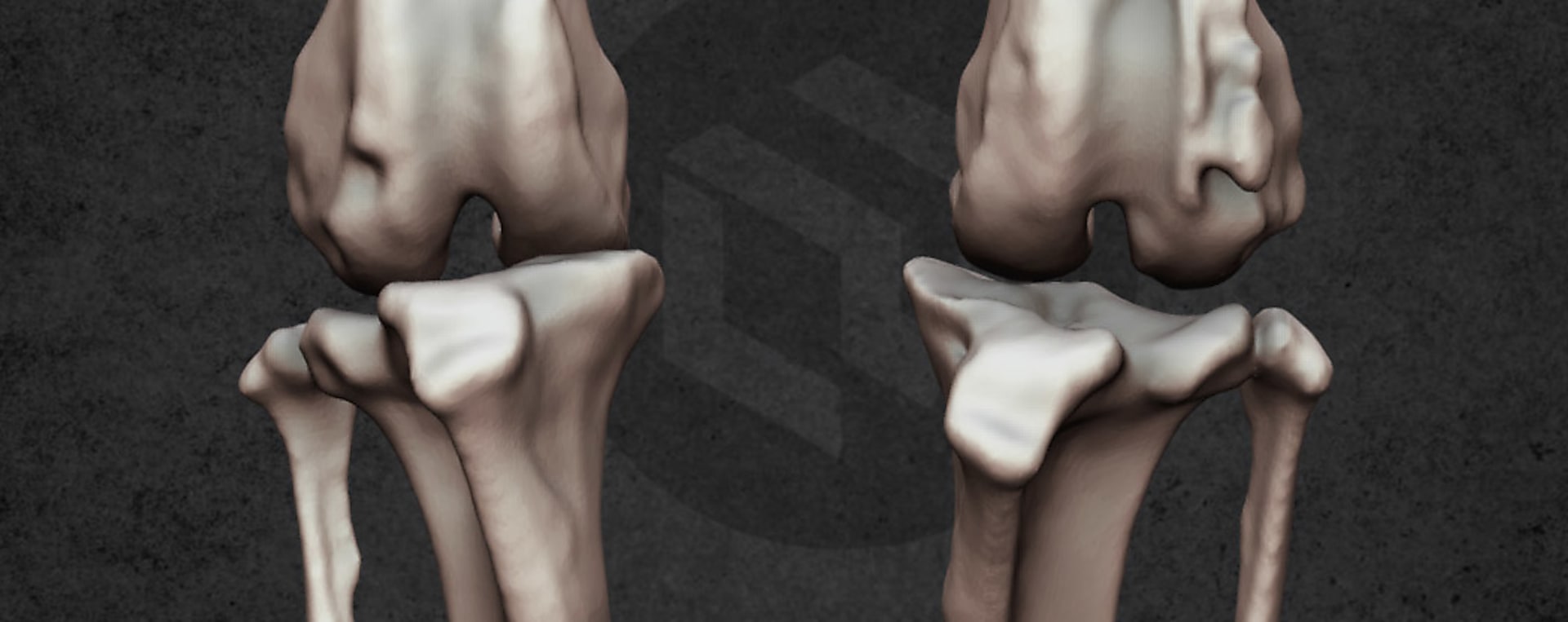
Pillar 4. Trochlear and Patellar Geometry: the tracking surface
Now we come to the groove - and it absolutely matters. The trochlea must be wide enough, deep enough, and have adequate ridge height to contain and guide the patella through movement. If the groove is shallow, dysplastic, or worn, surgical correction of that geometry is necessary.
Commonly applied techniques include trochlear wedge recession, block recession, and sulcoplasty - each with different implications for bone preservation, implant compatibility, and trochlear anatomy restoration. In some cases, patelloplasty is also relevant - particularly where patellar morphology, chronic wear, or incongruency compromises the articulation. Like all other steps, it forms part of the overall package rather than a standalone solution.
The most important reframe here is this:
The groove is the track.
Alignment is the steering.
You need both.
A perfectly deepened groove will not keep a patella on course if the limb's mechanical axis continues to push it sideways.
Section 5: Where customised sulcoplasty and trochlear implants fit
Customised trochlear solutions - implants designed to precisely restore or optimise the geometry of the sulcus - represent an important advance in managing trochlear dysplasia. Their value lies in the precision they offer: a surface-adaptive, congruent tracking surface that matches the patient's anatomy, with the potential to restore geometry with controlled bone preservation.
A customised groove solution is one highly controlled step in a multi-step process. Its outcomes depend directly on the quality of the surrounding plan:
- Has femoral or tibial malalignment been corrected where indicated?
- Has the tibial tuberosity been transposed to optimise the line of pull?
- Have soft tissues been balanced to support central tracking?
When those foundations are in place, a customised implant completes the solution elegantly. Without them, even a perfectly contoured groove will face mechanical forces it cannot overcome alone.
Section 6: The treatment planning flow
Bringing it all together, here is a structured checklist for approaching patellar luxation surgically:
- Confirm luxation pattern, grade, and functional impact;
- Evaluate full limb alignment - femoral and tibial axes, torsion, angular deformity;
- Decide whether deformity correction (angular and/or rotational osteotomy) is indicated;
- Realign the extensor mechanism - perform tibial tuberosity transposition where indicated;
- Balance soft tissues - release contracted structures, imbricate lax ones;
- Optimise trochlear and patellar geometry - sulcoplasty and/or patelloplasty as appropriate;
- Recheck tracking through the full range of motion under load where possible;
- Plan rehabilitation and post-operative monitoring.
Section 7: A conceptual contrast
Consider two cases - same breed, same grade of luxation, same shallow groove.
Case A: Groove deepening only. The trochlea is now geometrically better. But the femur still has internal torsion, the tibial tuberosity is still displaced medially, and the medial retinaculum is contracted. The patella tracks better for a few weeks - then gradually drifts back. Recurrence. Revision surgery.
Case B: Femoral torsion corrected. Tibial tuberosity transposed to restore a central line of pull. Soft tissues balanced - medial release, lateral imbrication. Groove geometry is then optimised with a customised implant that provides a congruent, adaptive tracking surface. The extensor mechanism now pulls straight, through a well-shaped groove, held by balanced soft tissues. Stable tracking. Good long-term outcome.
Same groove problem. Completely different result. The difference was the plan.
Take-Home Message
Patellar luxation is a whole-limb problem. Treating it well means thinking about three things equally.
The solution lives in three steps:
Correct the bones.
Control the soft tissues.
Contour the trochlea.
All three must be evaluated in every patient, and addressed in every case where they contribute to maltracking. Customised groove solutions are a powerful tool - most effective when integrated into a plan that has already accounted for the mechanics driving the problem upstream.
Thorough preoperative assessment, objective measurements, and technique selection matched to the individual patient's full limb mechanism are what separate good outcomes from great ones.
The groove is where the patella rides. The rest of the limb is what steers it there.
This article is intended for educational purposes and represents a treatment philosophy framework. Surgical decisions should always be made on a case-by-case basis by a qualified veterinary surgeon.