The Advantages of LimesVet Services for Your Veterinary Practice
Precise planning, safer surgeries! Integrate 3D technology into your veterinary practice and take advantage of the innovative solutions we offer.
Boci, a 5-year-old Beauceron dog, was suffering from severe neurological issues caused by spinal abnormalities. Dr. Csaba Pajor from the Váci Animal Hospital diagnosed the case. MRI and CT scans revealed that the last lumbar vertebra had fused with the sacrum, and spondylosis had developed between the last two lumbar vertebrae. Additionally, one of the intervertebral foramina had significantly narrowed, and the spinal cord was directly compressed by a bony protrusion extending into the spinal canal, resembling a small tooth. The surgery aimed to relieve the compression by removing the necessary parts while simultaneously stabilizing the affected vertebral bodies. At LimesVet, our task was to design a special surgical aid, known as a surgical guide, that helped identify the boundaries of the removable vertebral sections, including the internal tooth-like growth (exostosis), and determine the optimal locations and angles for the screw channels.
The surgery was successfully completed, and Boci's neurological problems were resolved.

Precise planning, safer surgeries! Integrate 3D technology into your veterinary practice and take advantage of the innovative solutions we offer.

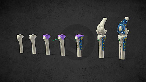
The combined application of custom 3D-printed and traditional implants represents a significant advancement in treating complex orthopedic conditions.

During the COVID-19 lockdowns, many people sought emotional support amid isolation and uncertainty. However, home office opened new opportunities for a young woman: finally, she could adopt a puppy. In the interview she talks about how thanks to this her life changed, how she found new social relationships and how her dog helped her maintain her mental health during this difficult period.
Patient-specific surgical equipment
Patient-specific surgical equipment
Patient-specific surgical equipment