3D Printed vs. Traditional Implants: Advantages, Challenges, and Innovative Solutions in Veterinary Care
While traditional implants have long been a reliable solution for various medical issues, 3D technology offers benefits that can significantly enhance the efficiency of surgical procedures and the recovery process for patients.
This article provides a detailed overview of how 3D-printed implants differ from standard implants, the advantages they offer, and the challenges faced when using them in veterinary practice.
Advantages of 3D-Printed Implants (3DI)
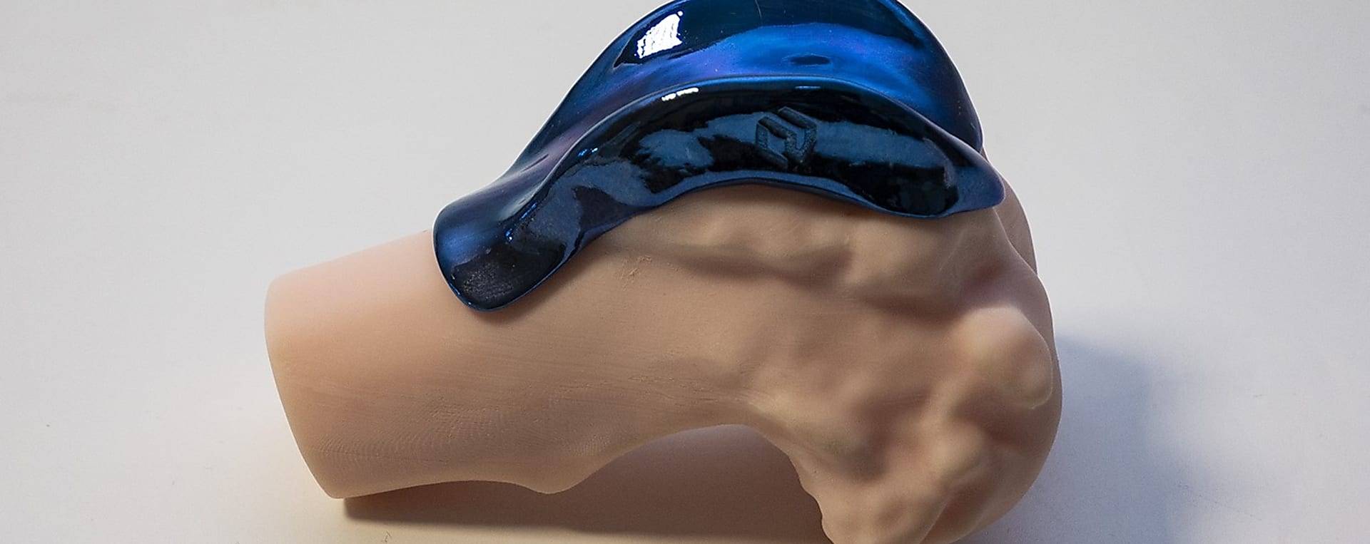
- Customization and Precision: 3D printing technology allows implants to be tailored to the specific anatomical features of the animal. The implants fit precisely, improving surgical outcomes and reducing the likelihood of postoperative complications.
- Cost-Effectiveness: Although the initial investment can be high, 3D-printed implants can be made available to smaller veterinary clinics through specialist providers.
- Innovative Solutions: 3D printing offers unique solutions such as implants with special surfaces that promote bone growth and the design of complex geometries that would be difficult to produce using traditional methods.
Areas for Improvement
- High Investment Costs: The acquisition of 3D printing equipment and necessary software can require significant investment. Additionally, the training and maintenance required to use the technology can be costly.
- Technical Complexity: Designing and producing 3D-printed implants requires high technical expertise, which can pose difficulties for smaller or less equipped veterinary clinics.
- Time-Consuming Design and Manufacturing: designing and producing custom implants can be time-consuming, which makes them less suitable for urgent cases, but this can be planned for in scheduled surgeries.
A Unique Solution Provided by 3D Technology When Traditional Implants Are “No Longer Enough”
A case study published in 20221 reported on a two-year-old Pomeranian dog experiencing severe issues 16 months after receiving a hip prosthesis.
Follow-up examinations revealed significant bone loss and medialization of the acetabular wall, which practically affected the dog's free movement.
Due to the patient’s small size, anatomical changes, and severe bone problems, traditional solutions were not feasible.
Instead, a custom 3D-printed titanium acetabular cage was created and implanted.
Two weeks post-surgery, almost completely normal limb functions were observed in the dog, and three years later, stable implant positioning and bone integration confirmed the success of the procedure.
Not Just Animals: 3D-Printed Implants Represent a Huge Leap Forward in Human Cases Too
Treating a failed ankle arthroplasty is a particularly challenging area; these cases are often addressed with TTC arthrodesis (fusion), which involves the fusion of the tibia, talus, and calcaneus using donor bone.
The problem is that these complex reconstructions often do not heal properly.
An example of this is a case study published in 20202, which details the story of a 65-year-old man.
In his case, ankle joint replacement was unsuccessful, and bone erosion and collapse were observed.
The solution was ultimately a 3D-printed titanium implant, which provided structural support to the affected area and facilitated bone integration, perfectly fitting the bones thanks to careful design.
Our Goal Is to Offer Modern Solutions in Veterinary Medicine
The continuous development of modern medicine and veterinary practice leads to new and innovative solutions, one of the most outstanding being the application of 3D printing technology in the field of implants.
LimesVet is committed to providing the most advanced technologies in veterinary care.
Our product range includes the latest 3D-printed implants, prostheses, and surgical guides, contributing to more efficient and safer surgical procedures.
References:
1A. Kang, H. Lee, Y. Roh, D. Kim, S. Jeong, J. Jeong (2022). Case report: Three-dimensionally printed patient-specific acetabular cage for revision surgery of aseptic loosening in a dog with micro total hip replacement. Frontiers in Veterinary Science. https://www.frontiersin.org/journals/veterinary-science/articles/10.3389/fvets.2022.915639/full
2R.J. Kadakia, N.B. Allen, A.E. Hanselman, S.B. Adams (2020). Clinical applications of custom 3D printed implants in complex lower extremity reconstruction. 3D Printing in Medicine. https://threedmedprint.biomedcentral.com/articles/10.1186/s41205-020-00083-4