TROCA 56
When severe patellar luxation requires precision — and planning makes all the difference
For them it is important to see objects in at least in 2D and to hold 3D objects in their hands to be able to understand and process information. This is the starting point of everything in the world of LimesVet as well.
Thanks to modern imaging technologies (such as CT and MRI), medical visualisation has become paramount because it can greatly help doctors make the right diagnosis. And 3D visualisation takes it even further. Nowadays there is a wide range of visualisation software and tools which enable professionals make well-founded decisions concerning the direction of the treatment.
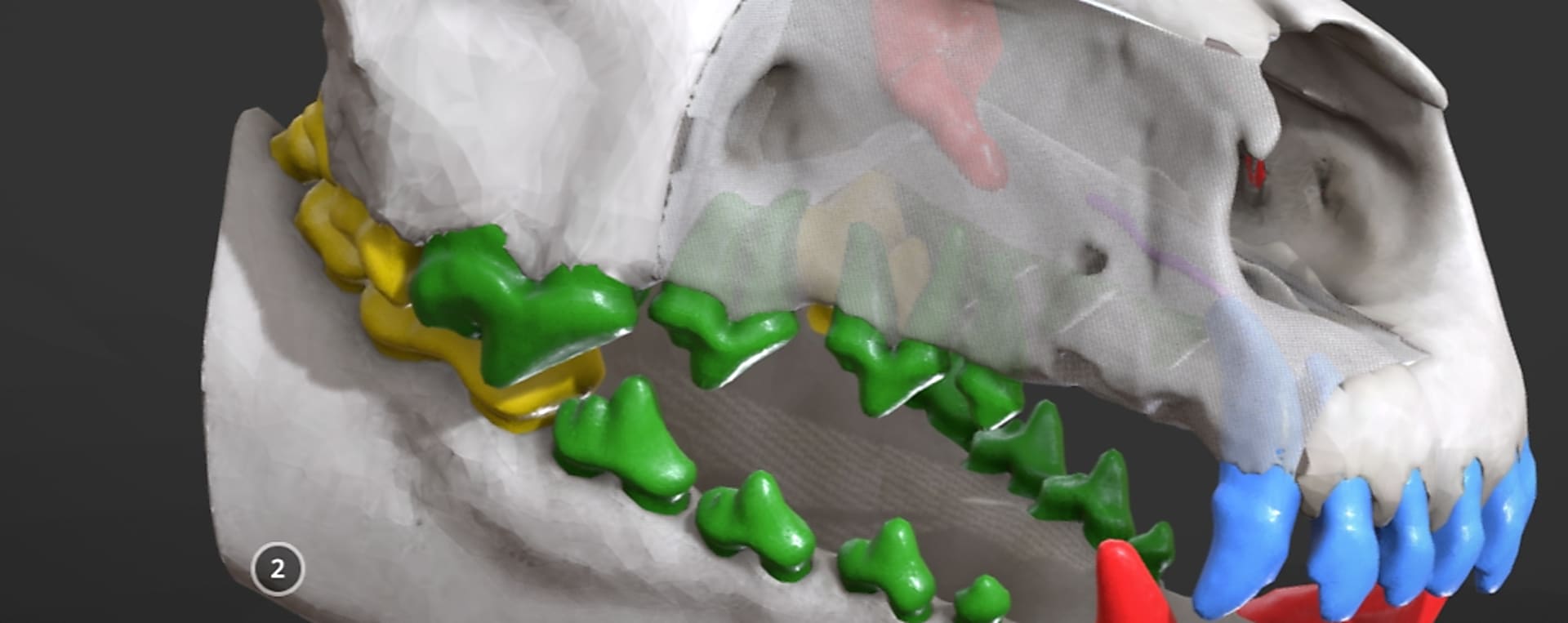
At LimesVet we strive to provide vets with as accurate models as possible. In the vast majority of cases, our work is based on CT or MR images from which we segment the relevant area modelled later in 3D. The models we extract in this way are worked out in detail with the help of various software. Besides, we also texture them depending on what we want to depict. The bones and anatomical structures can be coloured, made transparent or visualised in a lifelike manner. Then, therapists are provided with access to the model on an easy-to-use online interface where they can see and study the problematic area by rotating and zooming the model.
Why is it all useful? Because vets can see the area which needs treatment in a more lifelike and spectacular manner compared to CT and MR images without having to immerse into radiology. In addition, the more precise depiction makes the diagnostic process more efficient. One of our cases serves as an excellent example because the vet had to make a decision whether a cat can be operated or not. The lifelike 3D model made it clear that the owners make a better choice if they do not opt for surgery. It is also important to point out in connection with our cases that vets do not only have the opportunity to access the models but they can also consult with our experts to get a comprehensive picture based on which they can make a well-founded decision.
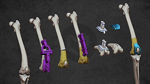
Of course, we have also had a lot of cases (in fact, these cases are in majority) when we could also contribute to operation planning. At this point we have to highlight the case when we had to assist with planning a complex operation by continuously cooperating with the surgeon in charge. With the help of the digital and 3D printed physical anatomical models the operation procedure was mapped out in detail enabling the vet to start the operation as prepared as possible and to carry out the process well and safely.
Now, we should turn our attention to the next level of visualisation, to the field of 3D printed anatomical models. The question arises as to why it is necessary to print the models if the digital model is at our disposal. At first glance, this question seems legitimate but, to our experience, physical models vastly support vets’ work. Their role is twofold because they make it possible for vets to visually explain different problems to pet owners and on the other hand, these models support the surgical planning process and, in some cases, the operation itself as well. The anatomical models can be printed in a scale of 1:1 or they can also be enlarged. They can be useful in the event of complex cases or by the preparation for operations when the surgeon has a small space to work in (e.g. in otolaryngology).
By applying anatomical visualisation we aim at providing vets with a high level of support as early as the stage of diagnosis. Besides, the prerequisite of using our other services is the creation of 3D models depicting the areas at issue.
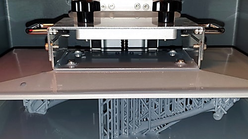
3D printing has an almost 40-year-long history of continuous development. Since its start, more and more printing technologies and materials have been emerging. We can create prints from hard filament, liquid resin and plastic or metal powder.

If you experience sudden muscle twitches or movement problems in your cat or dog, it can immediately scare you. Fortunately, however, small animal neurology is continuously developing which is greatly supported by our developments as well. In this episode of our interview series we are talking to Dr László Lehner, a veterinarian specialised in small animal neurology, who carries out numerous operations regarded as unique in Hungary, e.g. removing various brain tumours and implanting ventriculo-peritonealis shunts.

Diverse anatomical visualization
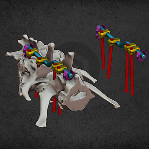
Patient-specific surgical equipment
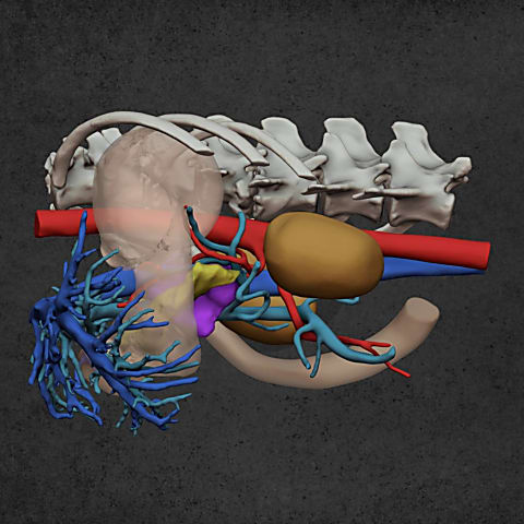
Diverse anatomical visualization
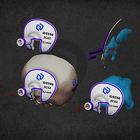
Patient-specific surgical equipment