

The Advantages of LimesVet Services for Your Veterinary Practice
Precise planning, safer surgeries! Integrate 3D technology into your veterinary practice and take advantage of the innovative solutions we offer.
This really interesting case arrived from a veterinary clinic in the countryside. Here the aim was to specify the diagnosis of a dental problem. First, we segmented the skull based on which we created a 3D model unambiguously showing the malformation of one of the dog’s teeth. His upper right canine tooth had not erupted in its proper place even after losing his deciduous tooth, so it stayed near the nasal cavity. From the perspective of visualisation we strived to show the teeth and the problem at the same time. Besides, we decomposed all these into steps so that the condition can be seen in multiple ways (by making the bones gradually transparent).
Precise planning, safer surgeries! Integrate 3D technology into your veterinary practice and take advantage of the innovative solutions we offer.
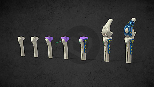
The combined application of custom 3D-printed and traditional implants represents a significant advancement in treating complex orthopedic conditions.
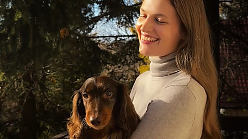
During the COVID-19 lockdowns, many people sought emotional support amid isolation and uncertainty. However, home office opened new opportunities for a young woman: finally, she could adopt a puppy. In the interview she talks about how thanks to this her life changed, how she found new social relationships and how her dog helped her maintain her mental health during this difficult period.
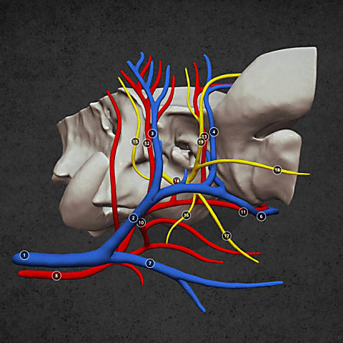
Diverse anatomical visualization

Diverse anatomical visualization

Diverse anatomical visualization