The Advantages of LimesVet Services for Your Veterinary Practice
Precise planning, safer surgeries! Integrate 3D technology into your veterinary practice and take advantage of the innovative solutions we offer.
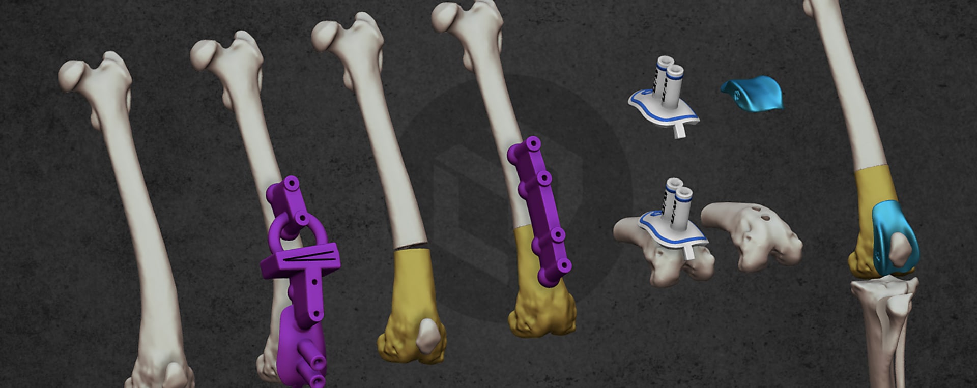
A mongrel dog was presented with grade IV medial patellar luxation in the left hind limb along with torsional limb deformity, a severe condition causing significant lameness, instability, and pain. In this case, correcting the trochlear groove alone was not enough - the femur and the tibia also required corrective alignment to restore proper joint mechanics.
To support the surgeon in this complex procedure, LimesVet was asked to deliver a solution that combined accurate correction with reliable implant placement.
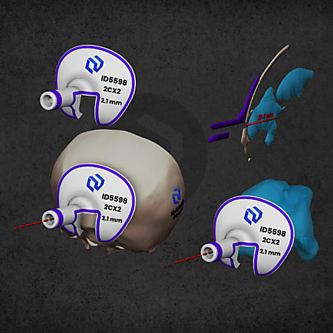
Using advanced 3D planning and patient-specific design, we created and 3D printed a custom surgical guide. This guide enabled precise femoral angular correction and helped define the exact positioning of the TROCA patellar groove prosthesis during surgery. In combination with the guide, the veterinarian used the TROCA 56 implant to restore proper trochlear surface and patellar route, and afterwards corrected the placement of the tibial tuberosity.
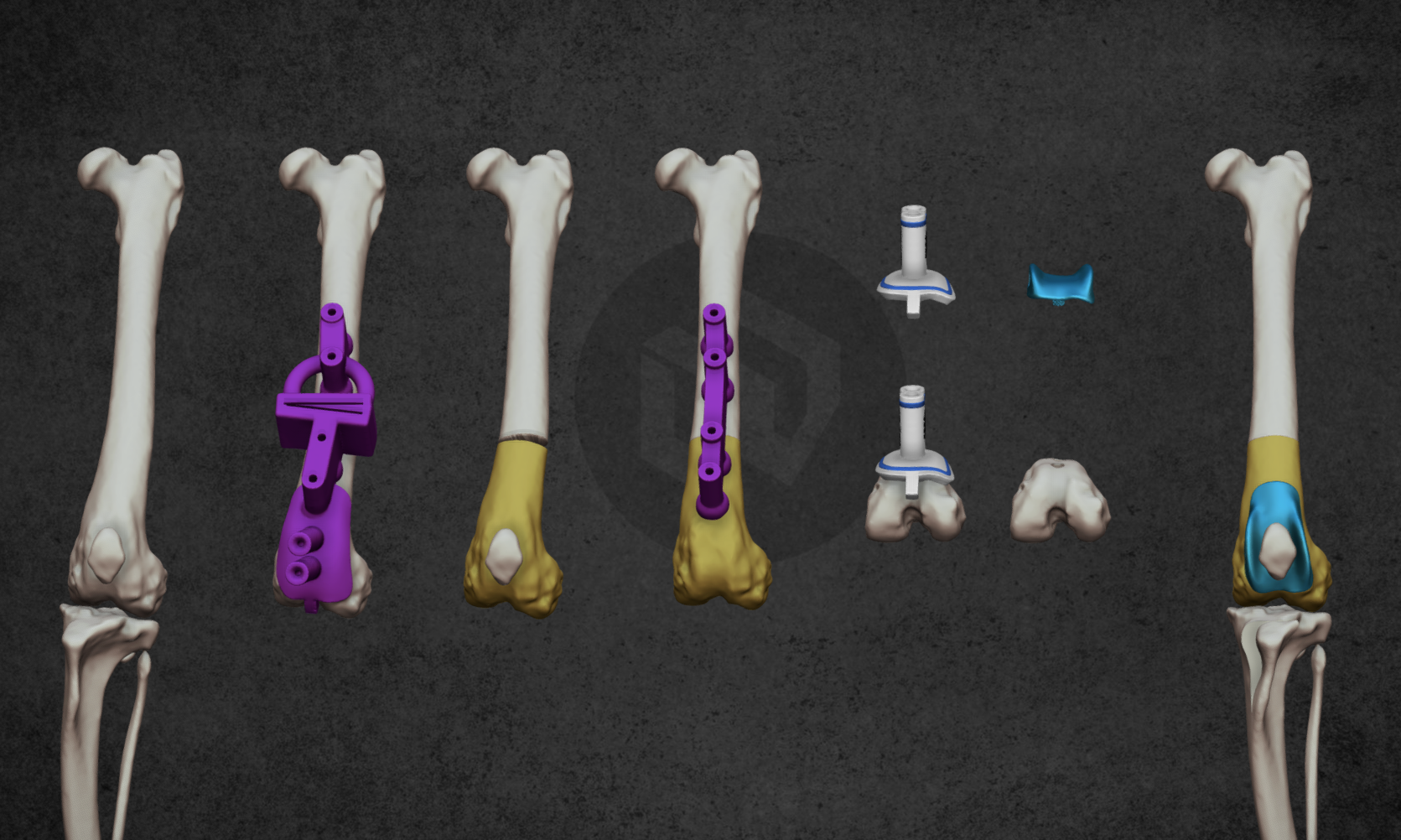
A key advantage of the TROCA system is that it does not require trochlear osteotomy of the patellar groove, while still fully matching the patient’s individual anatomy, and adjusting proper trochlear ridge heights. This allows for a less invasive approach, precise fit, and predictable biomechanical alignment.
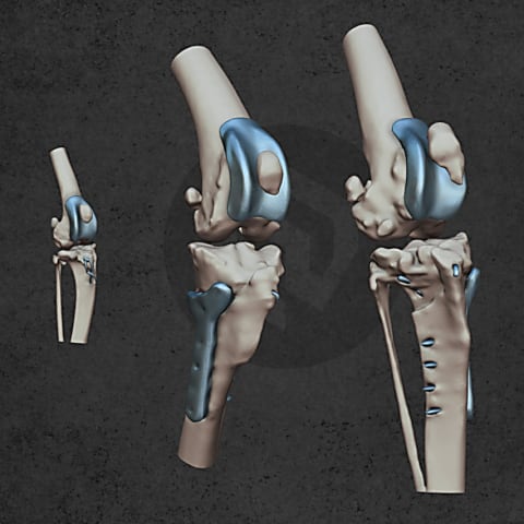
This integrated solution reduced intraoperative uncertainty, increased surgical confidence, and resulted in a more stable stifle joint and a smoother recovery for the patient.
Cases like this demonstrate how personalized 3D-printed surgical guides combined with anatomy-matching implant systems can elevate complex orthopedic procedures - delivering greater safety for the veterinarian and better outcomes for the patient.

Precise planning, safer surgeries! Integrate 3D technology into your veterinary practice and take advantage of the innovative solutions we offer.

Visualisation is actually as old as mankind. Even in prehistoric times people depicted animals in cave paintings, supposedly, to project a successful hunting. This form of art evolved approximately 30 thousand years before writing, so it can also explain why numerous people say that they are visual.

If you experience sudden muscle twitches or movement problems in your cat or dog, it can immediately scare you. Fortunately, however, small animal neurology is continuously developing which is greatly supported by our developments as well. In this episode of our interview series we are talking to Dr László Lehner, a veterinarian specialised in small animal neurology, who carries out numerous operations regarded as unique in Hungary, e.g. removing various brain tumours and implanting ventriculo-peritonealis shunts.
Personalized implants

Diverse anatomical visualization

Patient-specific surgical equipment
Patient-specific surgical equipment