TROCA 50
When chronic knee pain limits movement — and an anatomy-matched solution makes the difference

Virág was admitted to FuzioVet Animal Hospital for lameness examination because of the serious lameness of her left hind leg. Her condition was diagnosed as a serious limb malformation due to the congenital deformity of the left femur and the left tibia resulting in a fourth-degree medial patellar luxation - the inward dislocation of the patella. This was the cause of her lameness. The examinations made it clear that the traditional surgical solutions cannot achieve a state which could lead to a considerable improvement of the dog’s leg.
LimesVet got involved in the process at this point. The goal was to provide Virág with a custom designed, 3D printed stifle joint prosthesis which fits the faulty bones as much as possible. The following factors had to be considered and devised during the design process:
Virág’s case was immensely advantageous for LimesVet, too, because it was the first time we could make our entire range of services available, and so we could validate our processes. The whole development consisted of the following steps after determining the relevant factors:
1. Case study: had anyone prepared such prostheses for dogs? Result: we did not find any solutions in the world which could work in Virág’s case.
2. Clarifying anatomical questions: Our colleague, Kálmán’s deep knowledge of anatomy and Dr Antal Seregi’s professional experience and orthopaedic expertise clarified where, in what angle and how it is possible to cut the upper and the lower bones. We enhanced the efficiency of the analysis by creating digital 3D models of the bones which we also 3D printed so that we can also see the operation area by holding the models in our hands.
Result: the relevant parts of bones could be removed by keeping the muscles, and so not influencing the natural working mechanism of the leg (the important soft tissues, i.e. tendons, muscles, vessels and nerves were protected).
3. Specifying the working mechanism of the prosthesis, design process: First and foremost, the working mechanism of the prosthesis had to be specified because it served as the cornerstone of the further design process. On the one hand, the natural anatomical forms had to be restored. On the other hand, the attachment points between the prosthesis and the bones had to be planned as well. In addition, we applied a solution regarding the shape which makes it possible for the bone to grow into the prosthesis.
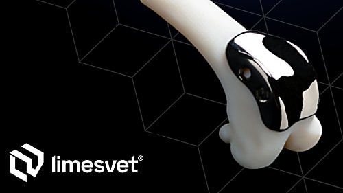
Result: we created the digital design of a single-axis stifle joint prosthesis which can easily be customised and manufactured.
4. Testing the shape and working mechanism by means of plastic printing: In this phase we 3D printed the bones and the prosthesis from plastic. By doing so, we could see whether the prosthesis fits the bones and how precise the 3D model is. With the help of the physical models we could also start planning the operation.
Result: the tests showed that the design process had been precise and that the attachments were adequate.
5. 3D printing of the prosthesis from surgical metal: big thanks to our strategic partner, Premet Ltd., because they have a such a great expertise in metal 3D printing and post-processing that the result was a prosthesis of excellent quality. Scinova Ltd. has to be mentioned, too, since they helped with manufacturing the drilled holes and the axis of the prosthesis.
Result: a high quality, 3D printed prosthesis of surgical metal (a special alloy of titanium) which works according to plan and which was customised to Virág.
6. The design and printing of special drilling and cutting guides necessary for a safe operation: As we are talking about a completely unique case, implanting the prosthesis also necessitates a unique solution. We designed custom-made cutting guides which we 3D printed from plastic so that the operation can be carried out according to plan and the bones and the prosthesis can be attached to each other appropriately.
Result: The guides performed well during the operation. By using them, Dr Seregi could cut the bones more safely and precisely at the required place, in the right direction and in the appropriate angle. The operation was successfully carried out and Virág’s healing process continued with the assistance of the experts at Ebfizio rehabilitation centre. Virág has been on rehabilitation since then but she already puts her weight on her leg and she started to use it. Virág’s story has a happy end as Dr Seregi took the dog home for the period of rehabilitation. Moreover, the family come to love her so much that they adopted her.
When chronic knee pain limits movement — and an anatomy-matched solution makes the difference
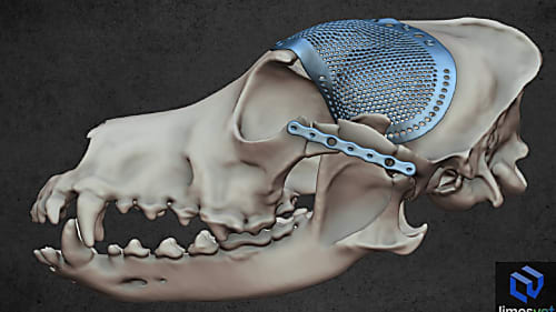

We are proud to have contributed as a partner to the success of a particularly complex cranial surgical case.
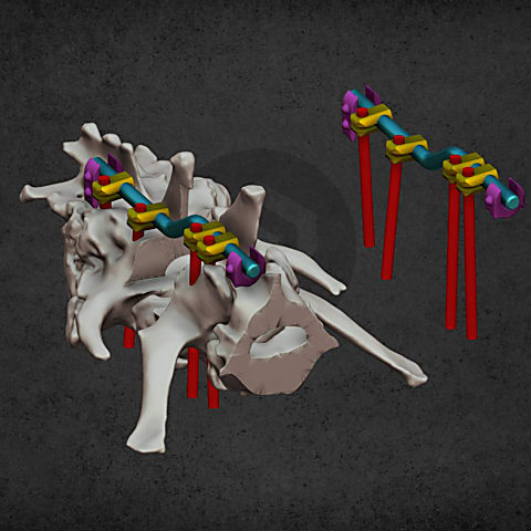
Patient-specific surgical equipment

Patient-specific surgical equipment

Patient-specific surgical equipment

Diverse anatomical visualization