Skullbase tumor

Skullbase tumor

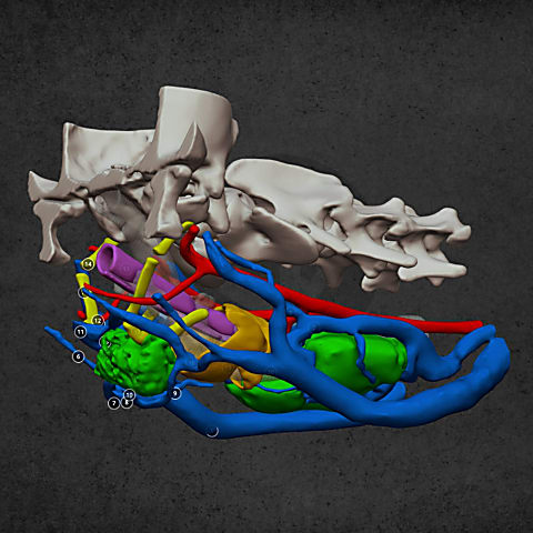
A CT scan in a Beagle dog revealed a tumor located at the base of the skull. The veterinarian requested that we use 3D visualization to show the major blood vessels in the surrounding area and assess whether surgical removal or reduction would be a realistic option. After careful evaluation of the case, the veterinarian decided to proceed with conservative therapy.

General
International Presence: Why We Build on Trade Shows

The role of 3D modeling in veterinary medicine is now undeniable, a fact clearly demonstrated by our experiences at leading European veterinary exhibitions.

General
The whole-limb puzzle behind patellar luxation

The misconception that leads to disappointing outcomes

References
Our products in action: a case where precision was critical

We are proud to have contributed as a partner to the success of a particularly complex cranial surgical case.

Skullbase tumor
Skullbase tumor

Diverse anatomical visualization

Hyoid tumor
Hyoid tumor

Diverse anatomical visualization

Dental visualisation

Diverse anatomical visualization

  Send case  Book a call