
The Advantages of LimesVet Services for Your Veterinary Practice
Precise planning, safer surgeries! Integrate 3D technology into your veterinary practice and take advantage of the innovative solutions we offer.
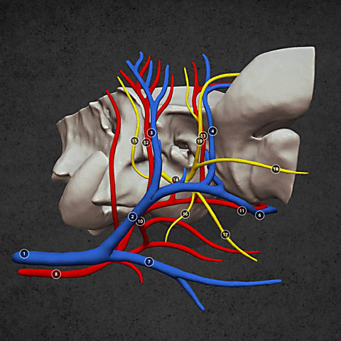
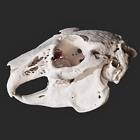
To fulfil a veterinarian’s request, we depicted a dog’s temporal bone in two ways. On the one hand, we visualised the main vessels and nerves running along the right external acoustic meatus on our model. On the other hand, we performed density analysis on the left temporal bone. The latter depiction can also show the spatial density of the bone based on the CT images, so in some cases it is more informative (due to its volumetric nature) compared to a 3D model concentrating solely on the boundary surfaces.

Precise planning, safer surgeries! Integrate 3D technology into your veterinary practice and take advantage of the innovative solutions we offer.
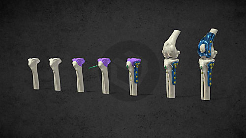
The combined application of custom 3D-printed and traditional implants represents a significant advancement in treating complex orthopedic conditions.

During the COVID-19 lockdowns, many people sought emotional support amid isolation and uncertainty. However, home office opened new opportunities for a young woman: finally, she could adopt a puppy. In the interview she talks about how thanks to this her life changed, how she found new social relationships and how her dog helped her maintain her mental health during this difficult period.
Diverse anatomical visualization
Diverse anatomical visualization

Diverse anatomical visualization