
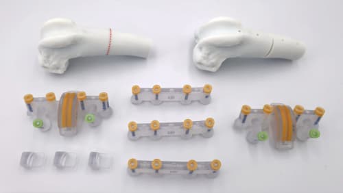
The FEMORISEC is a patient-specific, 3D-printed guide system for corrective femoral osteotomy in dogs and cats, most often distal femoral osteotomy (DFO). Includes CT-based cutting and repositioning guides.
FEMORISEC – Custom Femoral Osteotomy Guide | LimesVet
FEMORISEC is a patient-specific surgical guide system for corrective osteotomy of the femur in dogs and cats. It is designed from the patient's own CT scan and is most often used for distal femoral osteotomy (DFO), including cases where femoral deformity contributes to patellar luxation.
The role of femoral deformity
Angular deformities of the femur (such as distal femoral varus or valgus) and rotational deformities shift the line of pull of the quadriceps mechanism and change how the patella tracks. They are a frequent contributor to patellar luxation, and when they are left uncorrected, soft-tissue and trochlear procedures alone often do not give a lasting result.
Correction involves a wedge osteotomy at the right level and angle, followed by fixation of the segments in the corrected position. The procedure can be performed freehand; patient-specific guides help transfer the planned correction to the bone precisely and with consistent, reproducible accuracy from case to case.
How FEMORISEC works
We analyse the deformity in 3D, calculate the correction and design the patient-specific cutting and repositioning guides. You review and approve the plan before production. During surgery, the plan is transferred to the bone in two steps:
- Cutting guide - fits onto the femur in a single, predefined position, defines the location, planes and wedge geometry of the osteotomy, and sets the position of the reference K-wires.
- Repositioning guide - after the wedge is removed, it is placed onto the reference K-wires and holds the segments in the planned angular and rotational alignment until the plate is fixed.
This makes the correction predictable and consistent with the plan, with less intraoperative measuring and adjustment.
FEMORISEC and TROCA in one surgery
When a patient needs both deformity correction and trochlear resurfacing, FEMORISEC can be planned together with a TROCA 50 or TROCA 56 trochlear cap, using a combined guide. The DFO and the implant placement are then done in a single surgery, from one CT scan and one plan.
Process:
- Case submission: Attach a CT scan of the affected limb (DICOM format) and, if available, of the contralateral limb as a reference for comparison. CT requirements
- 3D visualisation: We create an accurate 3D model of the femur for your review within 2 business days.
- Planning and guide design: We analyse the deformity in 3D, calculate the correction and design the patient-specific cutting and repositioning guides. You review and approve the plan.
- Manufacturing: The guides and anatomical models are 3D printed from biocompatible material.
- Shipping: The complete kit is sent by courier and, with prompt approvals, usually arrives within 10 business days of case submission.
The FEMORISEC system includes:
- 1:1 anatomical bone models of the different stages (e.g. original and planned corrected state) for reference
- 2x patient-specific cutting guides - define the location, planes and wedge of the osteotomy
- 2x patient-specific repositioning guides - hold the segments in the corrected alignment until fixation
- All guides are labelled with drill and K-wire dimensions and a unique case identifier
- Sterilisation & handling instructions


