
Our products in action: a case where precision was critical
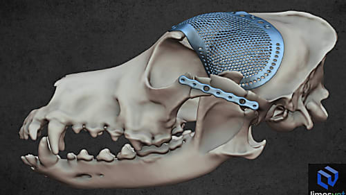
We are proud to have contributed as a partner to the success of a particularly complex cranial surgical case.
To fulfil a veterinarian’s request, we depicted a dog’s temporal bone in two ways. On the one hand, we visualised the main vessels and nerves running along the right external acoustic meatus on our model. On the other hand, we performed density analysis on the left temporal bone. The latter depiction can also show the spatial density of the bone based on the CT images, so in some cases it is more informative (due to its volumetric nature) compared to a 3D model concentrating solely on the boundary surfaces.

We are proud to have contributed as a partner to the success of a particularly complex cranial surgical case.
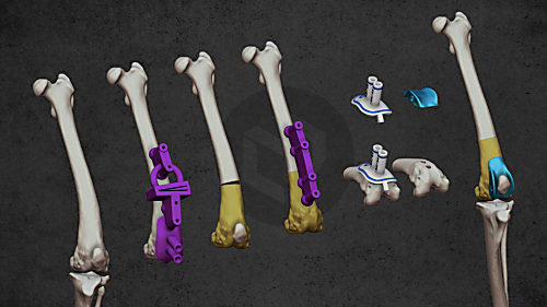
When severe patellar luxation requires precision — and planning makes all the difference
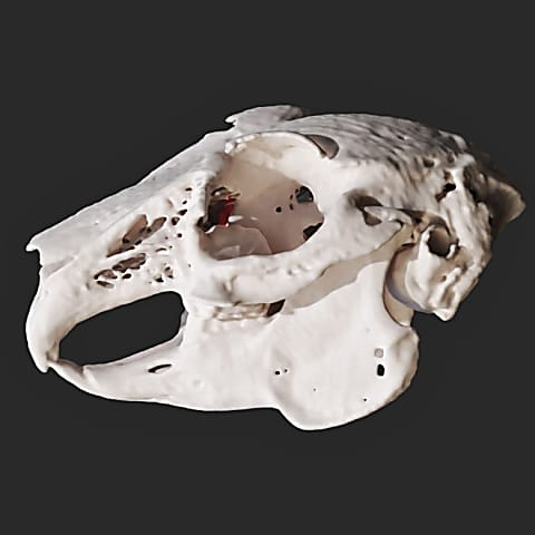
Diverse anatomical visualization

Diverse anatomical visualization

Diverse anatomical visualization