

The Advantages of LimesVet Services for Your Veterinary Practice
Precise planning, safer surgeries! Integrate 3D technology into your veterinary practice and take advantage of the innovative solutions we offer.
This case exemplifies very well how digital visualisation can help doctors see the problem adequately and make the right decision regarding operations. A vet sent us the CT image of a rabbit’s head for further analysis because the imaging examination detected a tumour in the back part of the nasal cavity near to the olfactory bulb. As he wanted to perform a minimally invasive surgical intervention, he asked us to create a 3D model making the position of the tumour more visible. We made the model and we could suggest an alternative surgical approach during the consultation.
Precise planning, safer surgeries! Integrate 3D technology into your veterinary practice and take advantage of the innovative solutions we offer.
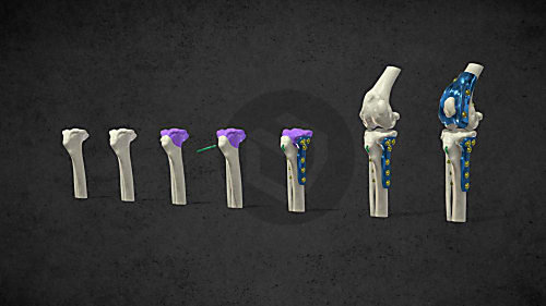
The combined application of custom 3D-printed and traditional implants represents a significant advancement in treating complex orthopedic conditions.
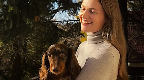
During the COVID-19 lockdowns, many people sought emotional support amid isolation and uncertainty. However, home office opened new opportunities for a young woman: finally, she could adopt a puppy. In the interview she talks about how thanks to this her life changed, how she found new social relationships and how her dog helped her maintain her mental health during this difficult period.

Diverse anatomical visualization

Diverse anatomical visualization

Diverse anatomical visualization