

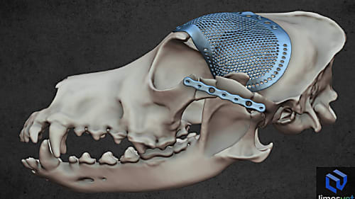
Our products in action: a case where precision was critical
We are proud to have contributed as a partner to the success of a particularly complex cranial surgical case.
This case exemplifies very well how digital visualisation can help doctors see the problem adequately and make the right decision regarding operations. A vet sent us the CT image of a rabbit’s head for further analysis because the imaging examination detected a tumour in the back part of the nasal cavity near to the olfactory bulb. As he wanted to perform a minimally invasive surgical intervention, he asked us to create a 3D model making the position of the tumour more visible. We made the model and we could suggest an alternative surgical approach during the consultation.

We are proud to have contributed as a partner to the success of a particularly complex cranial surgical case.
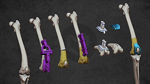
When severe patellar luxation requires precision — and planning makes all the difference
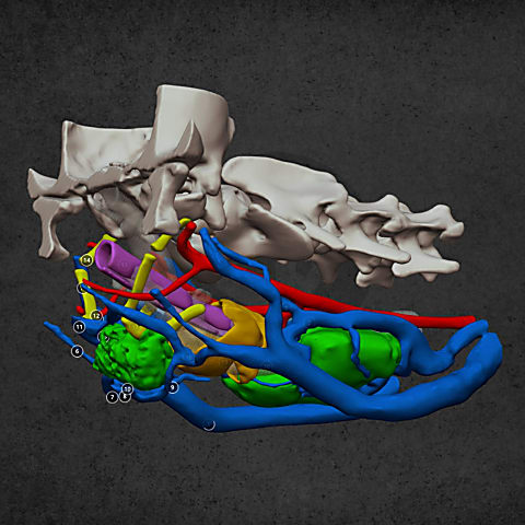
Diverse anatomical visualization

Diverse anatomical visualization

Diverse anatomical visualization