Our products in action: a case where precision was critical
We are proud to have contributed as a partner to the success of a particularly complex cranial surgical case.
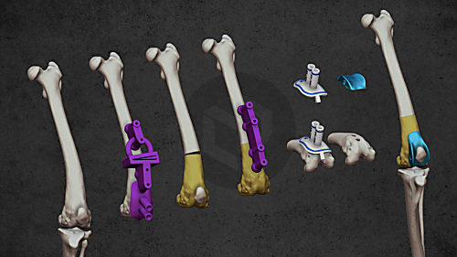
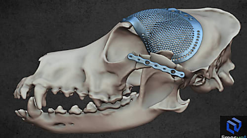
3D reconstruction of the skull and implant of a Miniature Pinscher who had a frontonasal squamous cell carcinoma. The fist models show how the tumor (which had soft tissue and osseous parts) was localised on the head, affecting the rfontal sinus, the medial wall of the orbit, and the inner and outer layer of the neurocranium. During the planning phase, as a safety margin was considered upon removal of the tumor, and a custom-designed titanium implant was made to cover the hiatus. The last model shows the 3D reconstruction from the postoperative CT scanning.

We are proud to have contributed as a partner to the success of a particularly complex cranial surgical case.

Personalized implants

Personalized implants

Personalized implants