
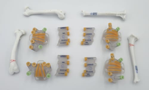
The TIBIOSEC is a patient-specific, 3D-printed guide system for corrective tibial osteotomy in dogs and cats with angular or rotational tibial deformities. Includes CT-based cutting and repositioning guides.
TIBIOSEC – Custom Tibial Osteotomy Guide | LimesVet
TIBIOSEC is a patient-specific surgical guide system for corrective osteotomy of the tibia in dogs and cats. It is designed from the patient's own CT scan and is the tibial member of LimesVet's osteotomy guide family, alongside RADISEC for the radius and FEMORISEC for the femur.
Tibial deformities and guided correction
Angular and rotational deformities of the tibia, such as tibial torsion or distal tibial varus, change the alignment of the whole pelvic limb. They load the stifle and tarsus abnormally, can cause lameness, and can contribute to patellar luxation.
Corrective tibial osteotomy can be performed freehand. Patient-specific guides add precision: the correction calculated on the 3D model is reproduced on the bone the same way every time, which supports consistent accuracy from case to case.
How TIBIOSEC works
Planning is supported by LimesVet's own 3D analysis method for tibial angular deformities. We analyse the deformity in 3D, calculate the correction and design the patient-specific cutting and repositioning guides. You review and approve the plan before production. During surgery, the plan is transferred to the bone in two steps:
- Cutting guide - fits onto the tibia in a single, predefined position, defines the location and planes of the osteotomy, and sets the position of the reference K-wires.
- Repositioning guide - after the cut, it is placed onto the reference K-wires and holds the segments in the planned angular and rotational alignment until the plate is fixed.
Part of whole-limb planning
Limb deformities often involve more than one bone segment. TIBIOSEC can be planned from the same CT scan as FEMORISEC and a TROCA trochlear cap, so femoral, tibial and trochlear corrections are designed as one consistent plan.
Process:
- Case submission: Attach a CT scan of the affected limb (DICOM format) and, if available, of the contralateral limb as a reference for comparison. CT requirements
- 3D visualisation: We create an accurate 3D model of the tibia for your review within 2 business days.
- Planning and guide design: We analyse the deformity in 3D, calculate the correction and design the patient-specific cutting and repositioning guides. You review and approve the plan.
- Manufacturing: The guides and anatomical models are 3D printed from biocompatible material.
- Shipping: The complete kit is sent by courier and, with prompt approvals, usually arrives within 10 business days of case submission.
The TIBIOSEC system includes:
- 1:1 anatomical bone models of the different stages (e.g. original and planned corrected state) for reference
- 2x patient-specific cutting guides - define the location and planes of the osteotomy
- 2x patient-specific repositioning guides - hold the segments in the corrected alignment until fixation
- All guides are labelled with drill and K-wire dimensions and a unique case identifier
- Sterilisation & handling instructions


