The Revolution of Surgical Guides in Veterinary Practice: 3 Case Studies Worth Reading
In the realm of veterinary operations, one of the biggest breakthroughs in recent years has been the advent of 3D-printed surgical guides.
These allow medical professionals to plan operations more precisely before the procedures, thereby reducing risks and increasing the success of interventions.
Numerous success stories testify to the significant advantages of 3D-printed guides in veterinary surgery.
Case studies confirm that these guides play a key role in reducing the number of complications and contribute to significantly shortening the healing time.
But to see how true this is, we can already present it through the description of a few specific cases:
A Three-Year-Old Pekingese Who Was Able to Walk Again
Bill Oxley's 2017 case study1 tells the story of a three-year-old male Pekingese who was unable to move independently due to the instability of his shoulders.
Thanks to computer-aided design programs and the use of patient-specific 3D-printed surgical guides, the stability of the shoulder joints was restored to a level where the dog was able to walk independently after two weeks, and later the normal movement without limping returned.
Tumor Removal with the Help of Surgical Guides
In the case of a 9-year-old female mixed-breed dog, symptoms of a meningeal tumor were observed, according to a 2022 case study2.
The dog had been suffering from regular seizures for three months, which, upon examination revealed a tumorous, cystic structure and cerebral edema on the MRI.
Prior to the surgery, patient-specific three-dimensional surgical guides were prepared using CT images and computer-aided 3D design programs to define the surgical approach, and the results spoke for themselves.
Thanks to the surgery, the tumor was completely removed, anticonvulsant therapy was suspended 90 days later, and the dosage of anticonvulsant drugs could be reduced in the second week.
225 days later, during the follow-up examination, it was confirmed that the seizures did not return, and the tumor did not recur.
4 Dogs Who Can Finally Move More Freely
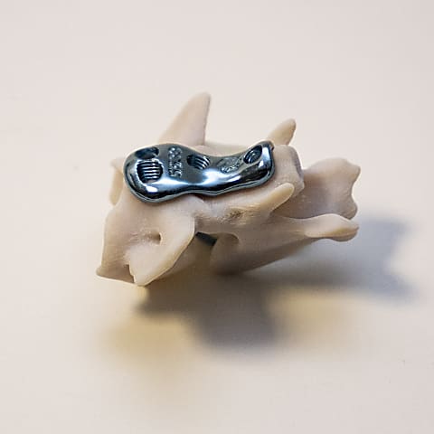
A study published in 2021 presents the treatment of limb deformities in not one, but four dogs, each case using 3D design and surgical guides3.
Based on the experiences, since the assistive devices could be specifically designed for the bones of the respective dog, there was no need for further measurements during the surgery: the restoration of the bones was accomplished more quickly and safely than in other cases.
Thanks to the interventions, in all four patient’s cases, it was possible to properly fix the affected legs without any complications.
Following the surgery, the owners of the dogs reported that their pets' main problem had been resolved, and they no longer stumbled over their front legs.
And These Are Just Three Examples among MANY
Entire books could be written about the studies and case descriptions published in professional journals in recent years on the successful use of 3D-designed surgical guides.
The above three stories are just a small taste of what the future holds in this area: the use of 3D-printed surgical guides revolutionizes veterinary surgery, increasing the accuracy of operations and reducing healing time.
As technology continues to evolve and veterinarians increasingly recognize its benefits, the use of 3D printed guides and other assistive devices is expected to become widespread – this is something we at LimesVet aim to further facilitate with our services and products.
References:
1Oxley, B. (2017). Bilateral shoulder arthrodesis in a Pekinese using three-dimensional printed patient-specific osteotomy and reduction guides. Veterinary and Comparative Orthopaedics and Traumatology: V.C.O.T, 30(3), 230–236. https://doi.org/10.3415/VCOT-16-10-0144
2So, J., Lee, H., Jeong, J., Forterre, F., & Roh, Y. (2022). Endoscopy-assisted resection of a sphenoid-wing meningioma using a 3D-printed patient-specific pointer in a dog: A case report. Frontiers in Veterinary Science, 9. https://doi.org/10.3389/fvets.2022.979290
3Carwardine, D. R., Gosling, M. J., Burton, N. J., O’Malley, F. L., & Parsons, K. J. (2021). Three-Dimensional-Printed Patient-Specific Osteotomy Guides, Repositioning Guides and Titanium Plates for Acute Correction of Antebrachial Limb Deformities in Dogs. Veterinary and Comparative Orthopaedics and Traumatology, 34(01), 043–052. https://doi.org/10.1055/s-0040-1709702


