

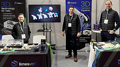
International Presence: Why We Build on Trade Shows
The role of 3D modeling in veterinary medicine is now undeniable, a fact clearly demonstrated by our experiences at leading European veterinary exhibitions.
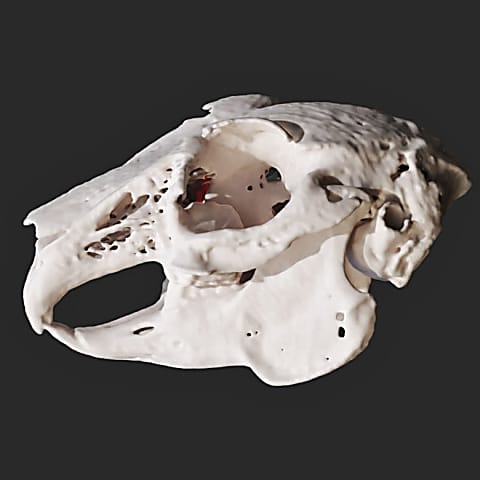
This really interesting case arrived from a veterinary clinic in the countryside. Here the aim was to specify the diagnosis of a dental problem. First, we segmented the skull based on which we created a 3D model unambiguously showing the malformation of one of the dog’s teeth. His upper right canine tooth had not erupted in its proper place even after losing his deciduous tooth, so it stayed near the nasal cavity. From the perspective of visualisation we strived to show the teeth and the problem at the same time. Besides, we decomposed all these into steps so that the condition can be seen in multiple ways (by making the bones gradually transparent).
The role of 3D modeling in veterinary medicine is now undeniable, a fact clearly demonstrated by our experiences at leading European veterinary exhibitions.

The misconception that leads to disappointing outcomes

We are proud to have contributed as a partner to the success of a particularly complex cranial surgical case.
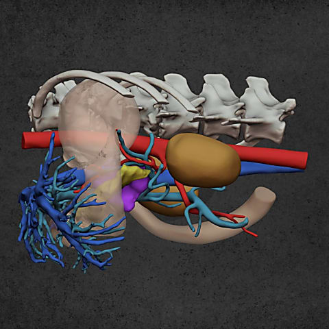
Diverse anatomical visualization
Diverse anatomical visualization

Diverse anatomical visualization