
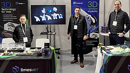
International Presence: Why We Build on Trade Shows
The role of 3D modeling in veterinary medicine is now undeniable, a fact clearly demonstrated by our experiences at leading European veterinary exhibitions.
To fulfil a veterinarian’s request, we depicted a dog’s temporal bone in two ways. On the one hand, we visualised the main vessels and nerves running along the right external acoustic meatus on our model. On the other hand, we performed density analysis on the left temporal bone. The latter depiction can also show the spatial density of the bone based on the CT images, so in some cases it is more informative (due to its volumetric nature) compared to a 3D model concentrating solely on the boundary surfaces.
The role of 3D modeling in veterinary medicine is now undeniable, a fact clearly demonstrated by our experiences at leading European veterinary exhibitions.

The misconception that leads to disappointing outcomes

We are proud to have contributed as a partner to the success of a particularly complex cranial surgical case.
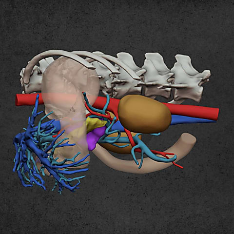
Diverse anatomical visualization
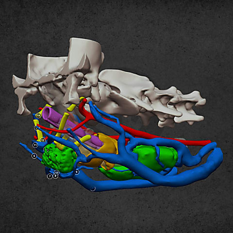
Diverse anatomical visualization

Diverse anatomical visualization