Ethmoidal tumour

Ethmoidal tumour

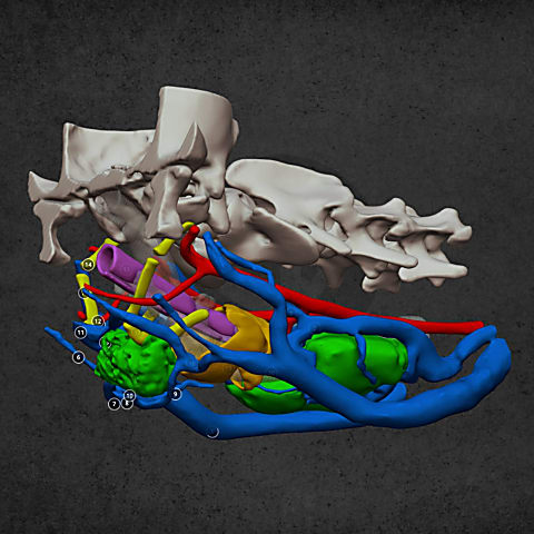
This case exemplifies very well how digital visualisation can help doctors see the problem adequately and make the right decision regarding operations. A vet sent us the CT image of a rabbit’s head for further analysis because the imaging examination detected a tumour in the back part of the nasal cavity near to the olfactory bulb. As he wanted to perform a minimally invasive surgical intervention, he asked us to create a 3D model making the position of the tumour more visible. We made the model and we could suggest an alternative surgical approach during the consultation.

General
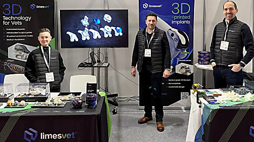
International Presence: Why We Build on Trade Shows

The role of 3D modeling in veterinary medicine is now undeniable, a fact clearly demonstrated by our experiences at leading European veterinary exhibitions.

General
The whole-limb puzzle behind patellar luxation

The misconception that leads to disappointing outcomes

References
Our products in action: a case where precision was critical

We are proud to have contributed as a partner to the success of a particularly complex cranial surgical case.

Palate defect
Palate defect

Diverse anatomical visualization

Pancreatic tumor
Pancreatic tumor

Diverse anatomical visualization

Ethmoidal tumour
Ethmoidal tumour

Diverse anatomical visualization

  Send case  Book a call