
International Presence: Why We Build on Trade Shows
The role of 3D modeling in veterinary medicine is now undeniable, a fact clearly demonstrated by our experiences at leading European veterinary exhibitions.
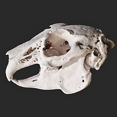
In this case a vet wanted to know the exact type and dimensions of the tissue hiatus the cat at issue had on its hard palate. These factors greatly determined the possibility of covering the hiatus and whether appropriate vascularisation can be ensured. In order for us to be able to provide the doctor with all the necessary information, we created the 3D model of the whole skull based on the CT images and we shared the zoomable and rotatable file with him.
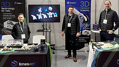
The role of 3D modeling in veterinary medicine is now undeniable, a fact clearly demonstrated by our experiences at leading European veterinary exhibitions.

The misconception that leads to disappointing outcomes

We are proud to have contributed as a partner to the success of a particularly complex cranial surgical case.
Diverse anatomical visualization
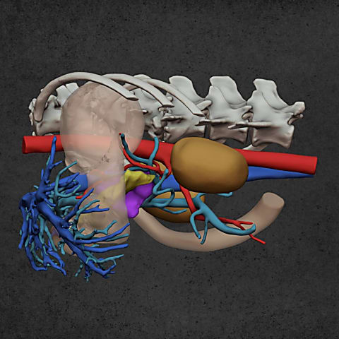
Diverse anatomical visualization
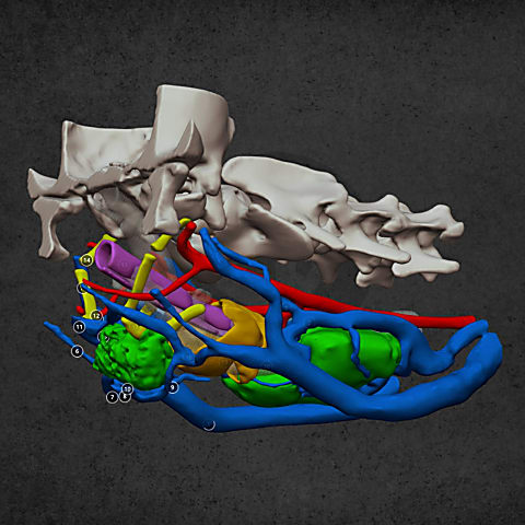
Diverse anatomical visualization